2024-12-27

The Variabilities of Dopamine (₯) - PART V: MeSH: D005239 & NBO:0000209 (Fear Comes in to Play)

Do you still remember the 67-year-old dopamine girl in the history of dopamine science this year? She gradually became a spokesperson for happiness in her twenties. However, behind this happiness is actually a little fear. In the early stages of research, scientists used basic tools to explore dopamine's role, focusing on its relevance to psychosis and antipsychotic drugs. Initial claims that dopamine was involved in fear conditioning were dismissed due to the inadequacies of the drugs, tools, and techniques used. However, with the development of science, it turns out that the purple "Fear" in "Inside Out" also has some relationship with the dopamine girl. Let's do some brain teasers together this time!
 
In "Dopamine at Forty," we learned that dopamine (DA) is more than just the "happy molecule" we once thought it was. Thanks to advancements in genetics, chemistry, and other technologies in the 21st century, scientists now understand dopamine and its interactions with neurons (DAN) and receptors much better.

These breakthroughs have shown us how dopamine is involved in both fear conditioning (how we learn to fear things) and fear extinction (how we stop fearing things). For example, a 40-year review of dopamine research revealed that different dopamine receptors play different roles in this process:
D1 in the amygdala “promotes” neuronal plasticity; D2 “inhibits” the plasticity of neurons; D3 in the ventral striatum (nucleus accumbens and olfactory tubercle) reflects functions such as fear, anxiety, and depression.
 
Figure 1: Dopamine, worker cells DAN and dopamine receptors are involved in fear conditioning and extinction
 
Canadian scientists, including Hamati, have studied dopamine and fear for almost 66 years. So why don't we hear much about dopamine's role in fear? Early research methods were limited, and most studies focused on animals, with human studies being relatively new. However, fear, like happiness, is one of our most basic emotions. By looking at Hamati's research, we can see which brain areas are active during fear conditioning and extinction. This new information about dopamine and its receptors is helping us understand the full story of dopamine's varied roles in the brain.
 
First, we first use the U.S. National Library of Medicine Medical Subject Title (MeSH) knowledge tree to quickly grasp fear (MeSH: D005239). The knowledge structure in behavior and behavioral mechanisms is shown in Figure 2. And the fear condictioning of Neuro Behavior Ontology (NBO) (NBO:0000209) is shown in Figure 3. According to the NBO's definition of fear conditioning: 
 A type of associative learning that allows organisms to acquire affective responses, such as fear, in situations where a particular context or stimulus is predictably elicits fear via an aversive context.
In other words, fear conditioning is the process by which our brains learn to associate certain things with fear. This mechanism plays a crucial role in how we respond to potentially threatening situations, where the brain makes connections between ordinary things and scary things.
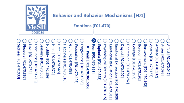
Figure 2: Fear MeSH: D005239 Knowledge Tree|Source: MeSH and revised by A.H. 
 Figure 3: Fear Conditioning: NBO: 0000209 knowledge tree | Source: NBO and revised by A.H.

Hamati et al. discussed the role of dopamine in classical fear conditioning and extinction. The process of encoding fear roughly includes several stages: Acquisition, Consolidation, Recall, Extinction(training, recall and return) of memory. In fact, what is most fascinating about DA's role in fear is its dual nature. While it helps encode fear, it also plays a role in the extinction of these memories. The study uses the acquisition and recall of conditioned fear stimulus (CS+) in classical conditioning theory to represent the ability to learn fear; the acquisition and recall of conditioned safety stimulus(CS−) as well as extinction training and extinction recall represent the ability to inhibit learned fear. Overall, both fear conditioning and extinction altered the activity of worker DAN cells and altered DA concentration levels in multiple brain regions, see Figure 4.
Figure 4: Effects of fear conditioning and extinction stages on DA levels in the brain|Source: adapted from Table 2 of Hamati et al.
 
  • Areas with increased DA activity levels: The amygdala is the most critical area in fear conditioning. DA increases during the acquisition and recall stages, along with terminal activity in the prefrontal cortex and most ventral midbrain periaqueductal gray matter (PAG)/ In the dorsal raphe nucleus (DRN) area, DA was increased.
  • Varies according to its location: Ventral tegmental area (VTA) in the animal literature suggests that DA is released in the medial prefrontal cortex (mPFC) during all stages of fear conditioning and extinction, but DAN activity in the medial and lateral VTA may depend on anatomy location and species. Because the number of active DANs in the substantia nigra pars compacta (SNpc) did not change after fear acquisition, the involvement of the SNpc in fear conditioning was not supported in the initial study. However, with the advancement of technology, during the fear acquisition process, the dorsolateral part of the SNpc has been suggested that regional DAN activity tends to increase, and the ventromedial region activity tends to decrease, which suggests that similar to the VTA, DA neuron activity in the SNpc varies depending on its location.
  • Uncertain region: Although the striatum, together with the amygdala, is considered a hub for coordinating fear responses and has the highest density of DA receptors relative to other brain regions, due to technical differences, the causal discussion on DA fear is still inconsistent and open for discussion.
Both D1-like and D2-like dopamine receptors help in learning and unlearning fear. D1-like receptors are important for picking up and solidifying memories, while D2-like receptors help in storing and recalling these memories. These receptors can work together in some brain areas or have their own unique roles. They work by sending signals through different brain regions like the amygdala, prefrontal cortex, hippocampus, and thalamus. See Figure 5.

Figure 5: Fear conditioning and extinction binding receptors|Source: adapted from Hamati et al. Figure 3
  • Amygdala and Prefrontal Cortex: Both these brain areas help in fear learning and unlearning. D1-like and D2-like receptors work together here. In the amygdala, when dopamine binds to these receptors, it helps create and stabilize fear memories. When recalling these memories, the brain reactivates them. On the other hand, getting rid of fear, like during fear extinction training, creates new circuits to suppress fear. In the medial prefrontal cortex (mPFC), dopamine helps inhibit fear, influenced by different subregions and types of neurons.
  • Hippocampus: The D1-like and D2-like receptors in the CA1 part of the hippocampus help in consolidating fear memories, recalling them, and unlearning them during extinction training.
  • Thalamus: Here, D2 receptors help in fear learning, while D1 receptors help in unlearning fear.
  • Olfactory Tubercle: This area, part of the nucleus accumbens, has neurons with dopamine receptors that respond to fear conditioning cues.
In the past, we thought of dopamine mainly as the chemical behind pleasure, motivation, and setting goals. But thanks to scientific progress, we now know dopamine does much more. Think of dopamine and the cells it works with (DAN) as storytellers, recording tales of curiosity, fear, and safety. Our brain isn't just a static memory bank; it's like a constantly changing landscape, shaped by our experiences. Dopamine sends signals through receptors that help form, strengthen, and retrieve memories of fear and how to overcome it.

Dopamine isn't just hanging out; it's a key player in learning fear. When we're conditioned to fear something, DAN cells light up, record fear memories, and send signals to parts of the brain like the amygdala and prefrontal cortex, which handle fear processing. Overcoming fear, or fear extinction, is like turning on a light in a dark room, slowly making the fear disappear. During this process, the brain learns that the once-feared thing is no longer a threat. Dopamine plays a dual role here, working with DAN cells and receptors to update and overwrite fear memories. Fear becomes a useful emotion, helping us prepare for danger. In the end, there's a balance between fear and safety, and dopamine helps maintain that balance.

Reference
  • Kawahata, I., Finkelstein, D. I., & Fukunaga, K. (2024). Dopamine D1–D5 Receptors in Brain Nuclei: Implications for Health and Disease. Receptors, 3(2), 155-181.
  • Hamati, Rami, et al. "65 years of research on dopamine's role in classical fear conditioning and extinction: A systematic review." European Journal of Neuroscience 59.6 (2024): 1099-1140

2024-12-25

The Variabilities of Dopamine (₯) - PART IV: MeSH:D011954

Dopamine is a pretty quirky character. Not only does it act as a neurotransmitter, but it also behaves differently depending on the "dopamine receptor" it binds to. Imagine these receptors as different doorways on the surface of a cell, each one changing how dopamine does its job. So, what's so special about these receptors? Well, think of them like the VIP passes that let dopamine into the cell club. You've probably heard about receptors because of the coronavirus (yep, the COVID-19 villain). The virus uses a special receptor called "ACE2" to sneak into our cells. Using this same idea, you can picture dopamine needing its own special receptors to get things done. In short, dopamine receptors are like the bouncers deciding who gets into the cell party, and without them, dopamine would just be left knocking on the door! 

Receptors on the surface of cell membranes are like the gatekeepers for cells to receive signals. These signals come from neurotransmitters, which are chemicals that help pass messages in the brain. When a neurotransmitter arrives, it interacts with G proteins in the cell membrane through special receptors on the surface, generating second messages inside the cell. These second messages kickstart various biochemical reactions in the cell, influencing downstream activities.

These receptors are called G protein-coupled receptors (GPCRs), the largest group of receptors that affect nearly all aspects of human physiology. Interestingly, about 40% of all approved drugs work by acting on GPCRs. Dopamine receptors are a type of GPCR found in neurons throughout the brain and body. When dopamine (DA) signals reach a nearby neuron, they bind to these specific receptors on the cell membrane of the neuron. This interaction helps regulate sensory and nerve signal transmission, as well as important processes related to cell balance and growth. 

Figure 1 shows how the small chemical molecule dopamine (DA) attaches to a larger dopamine receptor molecule and sends chemical signals by causing changes in the neurons that receive the signal. Think of the neurotransmitter (DA) and the receptor as a key and a lock. In this case, DA is the key and is called a ligand in biochemistry. A ligand is something that forms a complex with biological molecules to achieve a purpose. The word "ligand" comes from the Latin word "ligare," meaning "to bind." The ligand must fit well with its receptor, like a key fitting into a lock, to activate or inhibit protein activity, thus starting or stopping cellular responses. (part of the illustration of dopamine receptors uses BioRender components)

Next, let's dive into the expert definition of dopamine receptors.  

Figure 2 shows that, according to the Medical Subject Headings (MeSH) of the U.S. National Library of Medicine, the ID number for the dopamine receptor is D011954. It's defined as: “Cell-surface proteins that bind dopamine with high affinity and trigger intracellular changes influencing the behavior of cells.” From the MeSH dendrogram (a type of diagram showing relationships), we can see that the dopamine receptor is part of a larger protein family, which includes neurotransmitter receptors, biogenic amine receptors, and GPCRs. It's related to adrenergic receptors and has subcategories like D1 and D2 receptors, along with their subtypes D5, D3, and D4. 

      Receptors, Dopamine D1: A subfamily of G-PROTEIN-COUPLED RECEPTORS that bind the neurotransmitter DOPAMINE and modulate its effects. D1-class receptor genes lack INTRONS, and the receptors stimulate ADENYLYL CYCLASES.

  • Receptors, Dopamine D5: A subtype of dopamine D1 receptors that has higher affinity for DOPAMINE and differentially couples to GTP-BINDING PROTEINS.

      Receptors, Dopamine D2: A subfamily of G-PROTEIN-COUPLED RECEPTORS that bind the neurotransmitter DOPAMINE and modulate its effects. D2-class receptor genes contain INTRONS, and the receptors inhibit ADENYLYL CYCLASES.

  • Receptors, Dopamine D3: A subtype of dopamine D2 receptors that are highly expressed in the LIMBIC SYST­EM of the brain.
  • Receptors, Dopamine D4: A subtype of dopamine D2 receptors that has high affinity for the antipsychotic CLOZAPINE.

Simply put, dopamine receptors are divided into two different families based on their effects on adenylyl cyclase, an intracellular signal transduction and key enzyme involved in cell regulation: D1 and D2, or "D1 like receptors: D1, D5" and "D2 like receptors: D2, D3, D4", depending on whether their activation leads to stimulation or inhibition of the enzyme , and pharmacological differences.

Secondly, different descendant families are subdivided based on homology, pharmacological and biochemical properties. Endogenous dopamine or exogenous dopamine drugs activate receptors, also known as dopamine receptor agonists, and the activated protein activity can initiate cellular responses and increase dopaminergic activity in the brain, which helps It is used to reduce the symptoms of Parkinson's disease (dopamine deficiency); on the contrary, antagonists inhibit protein activity, shut down cell responses, and are used in schizophrenia with overactive dopamine.

In addition, dopamine receptors are also closely related to biological neuropsychological functions. In fact, the research on dopamine receptors was about 15 to 20 years behind the research on dopamine. The journal "Neuropsychopharmacology" mourned the late Canadian pharmacologist Philippe Philippe who was famous for his dopamine receptors. Philip Seeman mentioned that people's awareness of the existence of two dopamine receptor subtypes (later designated as D2) began with Professor Seeman's antipsychotic drug research in 1974. In 2024, scientists Kawahata and others from Tohoku University in Japan and the University of Melbourne in Australia reviewed nearly 40 years of research (1984~2023) and sorted out the impact of dopamine D1-D5 receptors in the brain on health and disease. They pointed out in the paper, biological functions of dopamine receptors extend beyond functions such as movement, cognitive memory, motivation, and drug addiction, revealing another potential reason why polypeptide dopamine is so variable. This study reviews the distribution of dopamine D1-D5 receptors, based on the physiological significance and disease correlation represented by different regions in the brain. It not only advances our basic knowledge of the central nervous system, but also provides powerful clues for therapeutic intervention.

In summary, the focus of Kawahata et al.'s research includes five major areas: striatum: ventral, dorsal, and nucleus accumbens; prefrontal cortex; subthalamic nucleus; limbic system: amygdala, hippocampus; midbrain : Substantia nigra pars compacta (SNc) and ventral tegmental area (VTA). The psychological and physiological functions of dopamine receptors D1 to D5 in Figure 3 include:

  • The "happy" function is mainly located in D2 in the ventral striatum.
  •   "Motivation and reward" are widely reflected in D1 to D4 of the striatum; D2 and D3 of the prefrontal cortex; D2 and D3 of the subthalamic nucleus; D3 of the amygdala; D2 of the substantia nigra and ventral tegmental areas D1 and D3. D4.

In addition, regarding the response to "novel things", last time we focused on the working cells dopaminergic neurons (DAN). This time we can combine dopamine receptors to more completely understand how brain neurons arouse curiosity. In response to novelty, D4 receptors may have the greatest contribution. "D4 curiosity" is mainly reflected in the dorsal striatum, prefrontal cortex, subthalamic nucleus and amygdala, and has a role in coping with novel things in the substantia nigra. 

Research on D4 receptors and their link to curiosity and novelty-seeking behavior is fairly recent, starting around 1996 (less than 30 years from now). Since studies vary based on the groups of people studied, methods used, and statistical tests applied, the findings may differ. To sum it up, dopamine receptors from D1 to D5 each have unique features, are distributed differently in the brain, and respond to dopamine in their own ways. 

Interestingly, more and more evidence suggests that different ways dopamine affects these receptors can influence our brains in surprising ways we didn't realize. Are you feeling curious about this now? Dopamine is doing its job! (to be continued)


 Reference

  1. 金克寧, 現代藥物標靶—G蛋白偶合受體之研究解析, 科學月刊. 516期. 2012.12.01
  2. 林書瑤,【化學奇境】受體‧受體‧受體, 台大科教中心CASE報科學, 2010.12.23
  3. wikipedia:ligand
  4. MeSH(D011954)
  5. Madras, B., George, S. In memoriam professor Philip Seeman (February 8, 1934-January 9, 2021). Neuropsychopharmacol. 46, 1229–1230 (2021).
  6. Kawahata, I., Finkelstein, D. I., & Fukunaga, K. (2024). Dopamine D1–D5 Receptors in Brain Nuclei: Implications for Health and Disease. Receptors, 3(2), 155-181. ; this study is limited to dopamine receptors in the central nervous system of the brain. Other non-central nervous system dopamine receptors including the heart, lungs, renal system, and pancreas are not discussed in this scope.
  7. Oak, J. N., Oldenhof, J., & Van Tol, H. H. (2000). The dopamine D4 receptor: one decade of research. European journal of pharmacology, 405(1-3), 303-327.; Paterson, A. D., Sunohara, G. A., & Kennedy, J. L. (1999). Dopamine D4 receptor gene: novelty or nonsense ? Neuropsychopharmacology, 21(1), 3-16.
  8. BioRender (2022). Distribution of Dopamine Neurotransmitters in the Human Brain.


2024-09-21

The Variabilities of Dopamine (₯) - PART III: CL:0000700 & SIO:000823

Fig 1: dopaminergic neurons (DAN) & curiosity
In the complex drama of the brain, there are many characters, but dopaminergic neurons (DAN) take the lead role. These neurons are always on the lookout for new things and solving puzzles, like a brainy Sherlock Holmes. Dopamine, the neurotransmitter, is their trusty sidekick, helping them stay curious and active.

But, like in any good story, there's a twist. Over time, these once-energetic neurons start to lose their zest for new experiences. This decline in curiosity mirrors our own aging. It raises an important question: what happens in the brain to cause this loss? What makes our inner Sherlock Holmes lose interest in the unknown?

Fig 2: underlying neural mechanisms of curiosity
Did you know? Inside your brain, there's a cell that looks like a thin ginseng root with branches that resemble ginseng whiskers. This fascinating cell is the dopaminergic neuron (DAN). When we see something new, it lights up the brain's circuits, making us want to explore, learn, and discover. DAN is like a detective in the brain, always drawn to new and unknown things, eager for excitement and new knowledge.

However, as we get older, this curiosity fades. Like a firework losing its spark, older people gradually lose interest in new things, both social and inanimate. Why is DAN so fascinated by new experiences? And as we age, what changes in DAN make it lose its curiosity?

It turns out that dopamine, a very versatile neurotransmitter, is not only linked to our happiness, desires, and goals but also closely connected to our "epistemic curiosity" and "empathic curiosity". As you read this, your brain is reacting, just like it would if you were really thirsty or hungry. The dopamine-sensitive areas in your midbrain light up—a phenomenon we call "epistemic curiosity." This curiosity drives us to explore and understand the world around us.

Fig 3: The knowledge tree of curiosity: SIO:000823

Another type of curiosity happens when we interact with other people. When we feel comfortable in social settings and try to understand what others are thinking and feeling, our brains release a lot of dopamine. This is called "empathic curiosity." Today, we are exploring how our curious detective, DAN, and dopamine play a role in triggering these two kinds of curiosity.

The Chinese version of Curiosity’s Knowledge Tree was added after the article was published on 2024/09/06. At the time of writing, popular science articles PART I and II based on the ontology hadn’t been published yet, so A.H. wasn’t sure if readers would like them. That’s why she wrote the articles on dopamine and curiosity using a traditional style. After Parts I and II were published, and readers loved them, she added information about curiosity by the Semanticscience Integrated Ontology (SIO) as shown in Figure 3. Curiosity is described as a positive emotion, related to desire.

To understand the connection, we need to look at dopamine, a chemical released by special neurons (called DAN) in the brain areas known as the ventral tegmental area (VTA) and substantia nigra pars compacta (SNc). Studies using brain scans have shown that high levels of curiosity and new experiences increase activity in these areas. A 2023 study published in “Communications Biology” (a sub-journal of “Nature”) titled “Reduction in the activity of VTA/SNc dopaminergic neurons underlies aging-related decline in novelty seeking. ” looked at the brain mechanisms behind curiosity. It explained why our interest in new things decreases as we age. Through experiments with mice, the study showed that the reduction in activity of VTA and SNc neurons is a key factor in the decline of curiosity due to aging.

This
Fig 4: experiment on social and inanimate curiosity
study used mice to explore the relationship between brain neurons and curiosity, showing how these neurons affect curiosity. Figure 4 shows the results of testing curiosity about social interactions (making new mouse friends) and inanimate objects (like shiny toys) in young mice and older rats. Scientists observed that young mice are more curious and explore these new experiences more frequently than older rats. Over time, young mice's interest in new friends increases, while older rats show stable but lower curiosity levels.

In general, older rats are less curious compared to young mice. Researchers measured and analyzed the activity of dopamine neurons in the brain, finding that older rats have less spontaneous neuron activity.

They also used specific drugs to control dopamine release in the brain. When they reduced dopamine activity in older rats, the rats became less curious. When they increased dopamine activity in young mice, the mice became more curious. The study concluded that the number of dopamine neurons decreases with age, leading to less dopamine and lower curiosity in older individuals. In humans, we lose about 6% of certain neurons every decade, which reduces dopamine availability.


In simple terms, young and healthy dopaminergic neurons (DAN) can send signals on their own, without needing extra input. This means they can regularly fire up spontaneously. As we get older, these neurons become less active and send fewer signals, which is linked to a decrease in curiosity. It's like the aging brain dims the lights of curiosity. However, by boosting the activity of these neurons and increasing dopamine release, we might be able to prevent or improve the decline in curiosity and thinking skills caused by aging or Alzheimer's disease.

In the 1950s, when Einstein was 63, he wrote to a friend saying, “People like you and I, though mortal of course like everyone else, do not grow old no matter how long we live...[We] never cease to stand like curious children before the great mystery into which we were born.” While few can keep Einstein's level of curiosity, which drives exploration and learning, we now know that curiosity is closely linked to the chemical dopamine and the curious DAN neurons.

With this new knowledge, maybe the detective in our brains won't retire early. Instead, we might grow old gracefully, keeping our curiosity alive. This understanding gives us hope that our inner detective will keep looking for new and interesting things, allowing us to age with wisdom and wonder.


Fig 5: Different representative signals of dopamine, in addition to happiness, desire, motivational goals, etc., there is also curiosity


REFERENCE

2024-08-01

The Variabilities of Dopamine (₯) - PART II: CL: 0000700

CellsAtWork:dopaminergic neuron (DAN, CL: 0000700)
 
Hi everyone, I’m Dopamine Girl. As an organic compound, I am what chemist ChEBI calls 18243. My relationship with you humans can start from the Cell Ontology ID number 0000700 defined by biologists. The working cell is a dopaminergic neuron (DAN, CL: 0000700), a neuron (cell) that uses dopamine as the neurotransmitter. Without further ado, let’s look at Figure 1 to understand DAN’s work characteristics and knowledge genealogy.

Learn about cells and dopamine from the Cell Ontology (CL), mainly because the cell knowledge described by CL is pretty mature and complete (20 years ago from 2004), covering cell types from prokaryotes to mammals, though not including plants cell. The rich diversity of CL can be seen from that it has imported 17 external ontologies and been reused by 86 external ontologies. CL is also widely used, such as the Human Cell Atlas (the Human Cell Atlas) established in 2016 by an international research alliance composed of more than 3,000 biologists, informatics scientists, and clinicians from 99 countries, and the Human Cell Atlas of the National Institutes of Health in the United States. The HuBMAP project uses CL as its core resource as well.

Fig 1: DAN in the Cell Ontology.

In the Variabilities of Dopamine (₯) - PART I:ChEBI:18243, we introduced the father-child relationship in ontology, which is the up-and-down relationship of the knowledge structure or the "depth" of the ontology. In fact, the expression of depth in CL can be as deep as 31 levels, which is the 31 generations of the family tree in vernacular. Figure 1 shows the direct upper layer of DAN is the neuron that releases catecholamines as neurotransmitters - catecholaminergic neurons, namely the father of DAN. They inherit some characteristics of the three major cell families of neural, secretory and electrically activities, and therefore help us to easily grasp the characteristics of DAN as a working cell that releases and secretes dopamine.

Second, we are generally familiar with DAN, which resides in the midbrain, so DAN here is the target tissue for the major neurodegenerative pathologies when it comes to Parkinson's disease. DAN has two lower subcategories in the CL description, among which the midbrain dopaminergic neurons are the children of DAN. Since DAN will degenerate or decrease with age or disease, etc., if the brain cannot produce enough dopamine, the work responsible for transmitting messages cannot be performed normally, which may easily lead to the deterioration of body movements, cognitive sensory emotions, or hormones, as well as mediation and other related diseases.

Another neuron that lives in the inner nuclear layer of the retina, the so-called dopamanergic interplexiform cell, is also a child of DAN. It is not common in the general introduction of dopamine, and related research is relatively little. His specialty is the use of dopamine to regulate nerves in the retina to adapt to light. If he cannot use enough dopamine, it may also cause visual impairment in Parkinson's patients.

Nevertheless, compared to DAN's father-son relationship, siblings of DAN (left-right relationship) are more complicated. The CL ontology describes more than 2,700 cell types, including 660 types of nerve cells and more than 540 types of neurons. Fortunately, we have another body gang to rescue. The U.S. National Library of Medicine's Medical Subject Headings (MeSH) is 70 years old (published since 1954). From the Figure 2, you can learn about the working cell DAN in the nervous and cellular system from the MeSH knowledge tree, and the overview of its neuron cell siblings.

Fig 2: DAN in the MeSH tree

In the ontology, the relationship between father, son, brother and sister is the basic "blood relationship", and another relationship including the description of external knowledge is also the solid power of the ontology. Just like writing a biography, after the three generations of ancestors and grandchildren have been explained, the highlight is to narrate the protagonist's experience and meaningful events. For example, the chemical entity ChEBI:18243 introduced before has simple inclusion, similarity and application relationships. In the second part, our protagonist is a living cellular entity - dopaminergic neuron (DAN), so "biological process" becomes an indispensable element for interpreting knowledge. Through the external gene knowledge ontology (GO) imported into CL ontology, we can further understand the daily working status of biological processes of cellular DAN (Figure 3)

Working cells DAN are neurons that release dopamine through synapses. According to the definition of synapse by the GO ontology: the connection between the axon of one neuron and the dendrite, muscle fiber or glial cell of another neuron.

    Fig 3: DAN, dopamine & synapse base on GO ontology
  • The junction between an axon of one neuron and a dendrite of another neuron, a muscle fiber or a glial cell.
  •  As the axon approaches the synapse it enlarges into a specialized structure, the presynaptic terminal bouton, which contains mitochondria and synaptic vesicles. At the tip of the terminal bouton is the presynaptic membrane; facing it, and separated from it by a minute cleft (the synaptic cleft) is a specialized area of membrane on the receiving cell, known as the postsynaptic membrane.
  • In response to the arrival of nerve impulses, the presynaptic terminal bouton secretes molecules of neurotransmitters into the synaptic cleft. These diffuse across the cleft and transmit the signal to the postsynaptic membrane.

The biological process of DAN (CL: 0000700) described by the cooperation of CL and GO ontology is shown in Figure 4. Its "relationship" or a grammatical metaphor for the relationship in the knowledge ontology is a verb:

  • has participant
  • has end location
  • results in acquisition of features of

The entities (metaphorically represented as nouns) involved in biological processes are:

  • Fig 4: DAN involved in biological processes, based on CL& GO ontologies 
    synaptic transmission, dopaminergic: The vesicular release of dopamine. from a presynapse, across a chemical synapse, the subsequent activation of dopamine receptors at the postsynapse of a target cell (neuron, muscle, or secretory cell) and the effects of this activation on the postsynaptic membrane potential and ionic composition of the postsynaptic cytosol. This process encompasses both spontaneous and evoked release of neurotransmitter and all parts of synaptic vesicle exocytosis. Evoked transmission starts with the arrival of an action potential at the presynapse.
  • dopaminergic neuron differentiation: The process in which a neuroblast acquires the specialized structural and functional features of a dopaminergic neuron, a neuron that secretes dopamine.
  • astrocyte-dopaminergic neuron signaling: Cell-cell signaling that mediates the transfer of information from an astrocyte to a dopaminergic neuron.

After learning some basic knowledge structure of dopamine as a chemical and working cells DAN, think about it again. If dopamine is multi-variable, then when DAN living in the midbrain releases dopamine, its impact on biology should undoubtedly be the same. Variable, and so on, what unexpected scientific commotion will dopaminergic neurons bring us then? (to be continued)


Reference